I was in a room with the National Programme Manager earlier this month. It wasn't a hotel room or a police cell, but it was both relaxing and arresting, and gave rise to some interesting questions. Not all of which have answers. Amongst these was the issue of whether or not we should consider using a single field of view for screening, rather than the two we currently employ, and having posed that question, Lynne Lacey stated that the HTA have reported very similar sensitivity and specificity for the detection of sight-threatening diabetic retinopathy (STDR) with just one 45º view.
When she said HTA, I'm assuming Lynne meant the Health Technology Assessment Programme and not the Horticultural Trades Association, although as we're talking about fields, I could be wrong. The former is part of the National Institute of Health Research, and "produces independent research information about the effectiveness, costs and broader impact of healthcare treatments and tests for those who plan, provide or receive care in the NHS". Which covers virtually all of us, and makes their views well worth a listen.
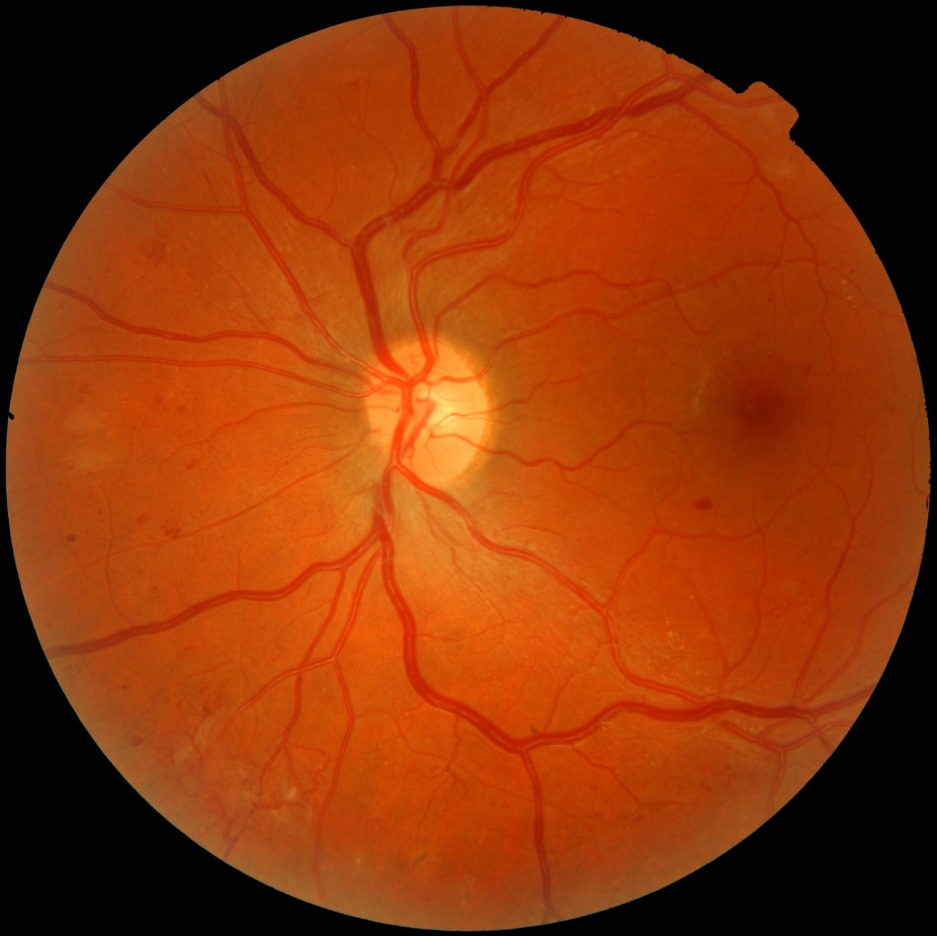
The NHS Diabetic Eye Screening Programme in England has always employed two 45º fields of view, one centred on the macula and the other on the optic disc, and to anyone familiar with this method, the suggestion of reducing our field of vision to a single photograph of each eye seems entirely counter-intuitive. Two images must be better than one. The more of the retina we can see, the more chance we have of spotting STDR, the more accurate our grading will be, and the less likely we are to miss something serious. We've all come across examples like this...

The temporal view shows, at worst, mild R2, but the nasal view reveals active new vessels. Relying on the first image alone would result in a routine referral, when an urgent one is clearly needed. With a bit of digging, I could probably find a macula-centred view which looks like a minor case of R1, complete with a disc-centred shot of R3. So it's case closed, surely? We need that second field.
Well, I'm not so certain. Back in 2004, the British Journal of Ophthalmology published this study from a team in Scotland which aimed to assess the effects of single versus three (rather than just two) field photography on screening for diabetic eye disease, and the results were somewhat startling. They concluded that using mydriasis and three field photography does not increase the sensitivity or specificity of detecting diabetic retinopathy. In other words, taking one photo without eye drops produces results which are just as accurate as those you'd get if you dilated the patient and took three photos of each eye. It might seem counter-intuitive, but it's peer-reviewed and in print.
Following on from this study, the Scottish Diabetic Retinopathy Screening Programme opted to go with single field photography, which it still employs to this day. In England we've always used two-field photography, but by putting this issue firmly on the agenda, Lynne Lacey has (quite rightly, in my opinion) started a debate which could have far-reaching consequences for the national programme. Put simply, this is a potential game-changer.
I spoke recently to a retinal screener from another programme who was telling me about the steady increase in her workload, and said that in order to meet the rising demand for screening without any increase in budget, she's being asked to screen almost forty patients a day. That includes measuring the visual acuity, dilating the pupils, recording any notes and taking the photographs. And she's not alone. Every screening programme in England is seeing its patient cohort grow year upon year, but I don't know of any with a rising budget to match. Programmes are consistently being asked to do more for less, and with that situation likely to continue, it's inevitable that the number of patients seen by each screener in clinic will have to go up.
As a retinal screener, that gives me concerns for my colleagues across the country, but equally I fear for the patients. The more people we see in each clinic, the less of a service they get. As patient numbers grow and appointment times shrink, the educational nature of the job will all but vanish, as the time to talk to patients becomes a luxury that no one can afford. Patients will end up being herded in and out of clinics by stressed, over-worked screeners who barely have the time to check a date of birth, never mind discuss a patient's condition. Photographs will be captured, but the quality of patient care will be diminished beyond recognition. All those added extras - the education, information and friendly, personal service that can transform a simple screening appointment into something far more valuable for the patient and just as rewarding for the screener - well, all of that will be lost.
Adopting a single field strategy would change that overnight. Taking only one photograph of each eye would dramatically reduce the need for drops, and with tropicamide priced at around 50p an ampoule, that would instantly slash costs. Removing the need for dilation, and halving the number of photos taken, would speed up the practicalities of the screening process and give the screener more time for patient education and feedback, while the likelihood of not having drops would encourage patients to attend, and reduce DNA rates. Meanwhile, back in the office, graders would have half the number of images to assess, and could potentially grade twice as many patients. Capacity would increase, as would patient uptake, and yet costs would realistically go down.
But what about the patient above? How do we manage the risk of an R3 patient masquerading as an R1 in a single temporal view?
Well, it's quite simple. We adopt a halfway house between single field and two-field photography. A few months ago, I mentioned that our national number-cruncher, Strat the Stat, had come to the conclusion that annual screening for patients with no retinopathy might be a waste of our resources. Well, so is two-field photography. And probably mydriasis too. I would implement a very simple new rule: all patients are subjected to single field retinal photography until such time as they have background retinopathy in both eyes. At that point, they're put on an annual rescreen and given two-field photography at future visits.
Adopting this rule would result in the overwhelming majority of patients being effectively screened with just a single photograph of each eye, whilst simultaneously ensuring that anyone at any risk of STDR gets a two-field screening. As technology advances, and cameras improve, more and more of those single field screenings will be possible without drops, and by limiting this method to those patients at lower risk, we can rest assured we're not missing anything, whilst successfully reducing costs.
Put it this way: in front of you is a patient with type 2 diabetes. They're diet-controlled, and received a grade of R0 at their last screening, twelve months ago. With your budget increasingly tight and your workload rising, do you really need to spend significant time and money dilating their pupils with tropicamide and capturing four distinct views in an unlikely search for sight-threatening diabetic retinopathy, or would one undilated photo of each eye suffice? I'd suggest that's one question which does have an answer.