I've always said that retinal screeners and failsafe are a lot like teenagers and sex: we all talk about it; no one really knows how to do it; but we think everyone else is doing it all the time, so we say we’re doing it too. And until BARS holds another Failsafe Discussion Day (or sex education class) that's likely to remain the case.
But one area where I always assumed the National Diabetic Eye Screening Programme differed from the average teenage boy was in the overwhelming compulsion to draw penises. Frankly I didn't think we had any (compulsion, not penises). Admittedly, I've been known to explain retinal images to patients with the words "If it looks like Mars, it's normal; if it looks like Venus, it's ischaemic; and if it looks like Uranus, it could be a macula hole", but I've never had the balls to mention male genitalia, and the only thing I've drawn on is my experience.
That all looks set to change, however, following the arrival of last month's Test & Training set.
Back in 2013, the journal Psychological Science published a study entitled The Invisible Gorilla Strikes Again, which was based on the phenomenon of 'Inattentional Blindness', whereby observers miss unexpected, but salient, events while engaged in other tasks. It had previously been demonstrated by videos such as this one:
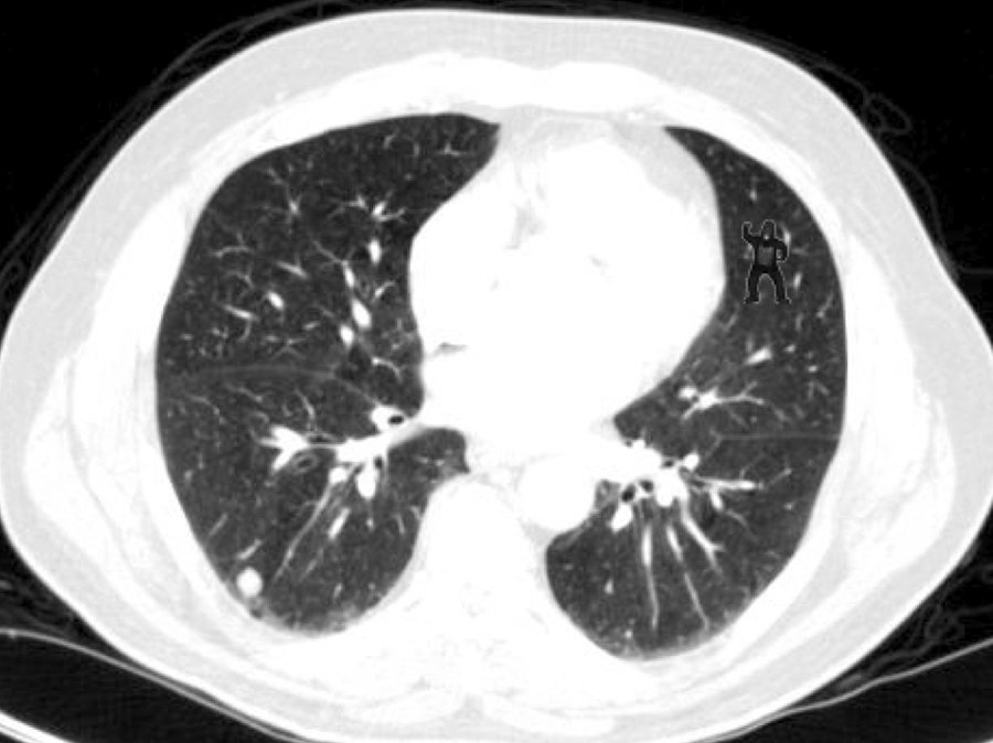
With the exception of Dian Fossey and the Harlem Globetrotters, however, most people viewing that video weren't specialists on the subject matter, so researchers at Harvard Medical School and Brigham & Women's Hospital in Boston decided to examine whether inattentional blindness also affects expert observers. They asked 24 radiologists to examine five CT scans of lungs for cancer nodules, a specialised task highly familiar to all of them. The last of those five scans looked like this...

Despite being highly skilled observers, 20 of those 24 radiologists, or 83%, failed to spot the gorilla in the top right hand corner. The other 4 went ape when they saw it.
The results indicate not that the radiologists weren't looking carefully enough, but rather that their brains were so focused on spotting cancer nodules, they were blind to anything else.
The resulting press coverage of this study prompted a lot of discussion in my screening programme about whether or not we would notice such a thing when grading a retinal image. Most graders found it hard to believe they could ever miss anything so out of place. But then the radiologists probably would have said the same thing too. And let's face it, you could hide Lord Lucan in a nasal left view being graded after four-thirty on a Friday afternoon, and we'd all be none the wiser.
My conclusion was that there could be no better environment to further test this phenomenon than a diabetic eye screening programme. Not only would we be reducing the risk of blindness, we'd be assessing the risk of inattentional blindness. Once you include the blind luck required to get 100% on the TAT, we'd be on the verge of designing a triple threat study of high quality sight-impaired research, straddling Diabetes Care, The British Journal of Ophthalmology and Psychobabble Weekly.
And if I was thinking that way, I felt sure the national team were too. For the past two years I've been quietly confident that somewhere in Gloucester, behind closed doors, in a dimly lit room, and possibly in the dead of night, someone was green-lighting a top-secret research project to test the inattentional blindness of screeners, and that one day I would open an apparently innocuous image on the TAT, only to be confronted with a picture of Steve Aldington, the silverbacked godfather of grading, beating his chest as he swings through the temporaral arcade, grabbing veins like vines and shaking his fist at the camera.
Sadly it hasn't happened. Although if it had, 83% of us wouldn't know.
The thing about blind faith, however, is that it's eventually rewarded with a sight worth savouring. The April Test & Training set contained an unusual error in screen 005, which was assigned a ground-truthed grade of R0M0, despite the clear presence of retinopathy. My assertion is that this was a deliberate and subtle clue to the following month's invisible gorilla warfare...
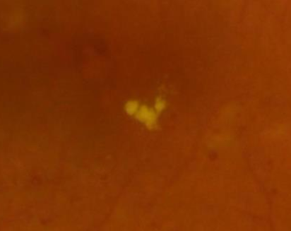
Screen 035 in May's Test & Training set was this temporal left image, graded as R2M1:

It's clearly M1. But in this case the M stands for Member...

So there you have it. The April set featured a cock-up. The May set featured a cock. And I'm standing proud amongst the 17% who saw it.

